 국립중앙도서관 "우편 복사 서비스"로 연결 됩니다.

 ScienceON
Purpose : To evaluate the extent of tissue coagulation during interstitial laser photocoagulation (ILP) innormal bovine liver, using a diode laser unit and various parameters, and to determine whether the procedure isapplicable to clinical practice.. ...
다국어 입력
あ
ぁ
か
が
さ
ざ
た
だ
な
は
ば
ぱ
ま
や
ゃ
ら
わ
ゎ
ん
い
ぃ
き
ぎ
し
じ
ち
ぢ
に
ひ
び
ぴ
み
り
う
ぅ
く
ぐ
す
ず
つ
づ
っ
ぬ
ふ
ぶ
ぷ
む
ゆ
ゅ
る
え
ぇ
け
げ
せ
ぜ
て
で
ね
へ
べ
ぺ
め
れ
お
ぉ
こ
ご
そ
ぞ
と
ど
の
ほ
ぼ
ぽ
も
よ
ょ
ろ
を
ア
ァ
カ
サ
ザ
タ
ダ
ナ
ハ
バ
パ
マ
ヤ
ャ
ラ
ワ
ヮ
ン
イ
ィ
キ
ギ
シ
ジ
チ
ヂ
ニ
ヒ
ビ
ピ
ミ
リ
ウ
ゥ
ク
グ
ス
ズ
ツ
ヅ
ッ
ヌ
フ
ブ
プ
ム
ユ
ュ
ル
エ
ェ
ケ
ゲ
セ
ゼ
テ
デ
ヘ
ベ
ペ
メ
レ
オ
ォ
コ
ゴ
ソ
ゾ
ト
ド
ノ
ホ
ボ
ポ
モ
ヨ
ョ
ロ
ヲ
―
http://chineseinput.net/에서 pinyin(병음)방식으로 중국어를 변환할 수 있습니다.
변환된 중국어를 복사하여 사용하시면 됩니다.
예시)
- 中文 을 입력하시려면 zhongwen을 입력하시고 space를누르시면됩니다.
- 北京 을 입력하시려면 beijing을 입력하시고 space를 누르시면 됩니다.
А
Б
В
Г
Д
Е
Ё
Ж
З
И
Й
К
Л
М
Н
О
П
Р
С
Т
У
Ф
Х
Ц
Ч
Ш
Щ
Ъ
Ы
Ь
Э
Ю
Я
а
б
в
г
д
е
ё
ж
з
и
й
к
л
м
н
о
п
р
с
т
у
ф
х
ц
ч
ш
щ
ъ
ы
ь
э
ю
я
′
″
℃
Å
¢
£
¥
¤
℉
‰
$
%
F
₩
㎕
㎖
㎗
ℓ
㎘
㏄
㎣
㎤
㎥
㎦
㎙
㎚
㎛
㎜
㎝
㎞
㎟
㎠
㎡
㎢
㏊
㎍
㎎
㎏
㏏
㎈
㎉
㏈
㎧
㎨
㎰
㎱
㎲
㎳
㎴
㎵
㎶
㎷
㎸
㎹
㎀
㎁
㎂
㎃
㎄
㎺
㎻
㎽
㎾
㎿
㎐
㎑
㎒
㎓
㎔
Ω
㏀
㏁
㎊
㎋
㎌
㏖
㏅
㎭
㎮
㎯
㏛
㎩
㎪
㎫
㎬
㏝
㏐
㏓
㏃
㏉
㏜
㏆

RISS 인기검색어


소 간에서 다이오드레이저를 이용한 간질성 광응고술 = Interstitial laser photocoagulation with diode laser unit in bovine liver
한글로보기https://www.riss.kr/link?id=A100883721
- 저자
- 발행기관
- 학술지명
- 권호사항
-
발행연도
1999
-
작성언어
Korean
- 주제어
-
등재정보
KCI등재,SCOPUS
-
자료형태
학술저널
- 발행기관 URL
-
수록면
39-45(7쪽)
- 제공처
-
0
상세조회 -
0
다운로드
부가정보
다국어 초록 (Multilingual Abstract)
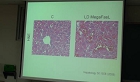
Purpose : To evaluate the extent of tissue coagulation during interstitial laser photocoagulation (ILP) innormal bovine liver, using a diode laser unit and various parameters, and to determine whether the procedure isapplicable to clinical practice.. Materials and Methods : Using an 18-gauge needle, experimental interstitiallaser photocoagulation (ILP) was carried out in normal bovine liver. On the basis of differing parameters, threegroups were established. For groupI, a single photofiber with laser power of 1, 3 and 5 watts and an exposuretime of 60, 180, 300, 420 or 600 seconds was used. For groups II and III, four needles were fixed at a distance of1cm and 1.5cm ; in each case a needle fixation device was used, as well as a laser distributor for simultaneouslaser exposure of photofibers. As a control, four photofibers were placed as for groupIII, but to compare groups IIand III, each photofiber was exposed to a laser of 3 watts 300 seconds, without using a laser distributor. Toevaluate the range of tissue coagulation, specimens were analyzed both with regard to cross-sectional grossfindings and histopathologically . Results : The largest diameter of thermal coagulation necrosis in GroupI was15$\times$15mm, and this was ball-shaped. Coalescence of coagulation between each photofiber was observed in GroupII,and this was up to 25 mm in diameter. In GroupIII and controls, coalescence was not found, though the extent oftissue coagulation increased with increasing wattage and exposure time. The extent of charring at the center ofcoagulation also increased with increasing wattage. Smoke bubbles emanating from the coagulation area wereobserved, and during ILP involving a single photofiber, increased from 3 watts, applied for 300 seconds.Conclusion : Using an 8-gauge needle and a diode laser ILP, we have shown that a range of tissue coagulationacutely ablates normal bovine liver. In selective cases, the procedure could be applied to clinical trials.
동일학술지(권/호) 다른 논문
-
대동맥류에 대한 Gianturco 스텐트와 PTFE를 이용한 스텐트-그라프트의 임상적 응용
- 대한영상의학회
- 박재형
- 1999
- KCI등재,SCOPUS
-
조영제 분할 주입법을 사용한 전체 대동맥과 장골동맥의 나선식 CT 혈관촬영술 : 고식적 단상 조영제 주입법과 비교 연구
- 대한영상의학회
- 편래현
- 1999
- KCI등재,SCOPUS
-
폐암의 종격동 림프절 전이 평가에서 전산화단층촬영의 정확도 : 전향적 연구
- 대한영상의학회
- 김영한
- 1999
- KCI등재,SCOPUS
-
범발성 간질성폐렴에서 간유리음영의 추적 고해상 CT 소견
- 대한영상의학회
- 김형환
- 1999
- KCI등재,SCOPUS
분석정보
이 자료와 함께 이용한 RISS 자료
나만을 위한 추천자료


